Forearm Fractures
Fracture Treatment at Children’s Mercy
Forearm fractures are the most common fractures in children, often occurring from a fall while playing and running. Because children’s bones are still growing, most of these fractures can be treated without surgery by means of splints or casts. Occasionally, they require reduction (repositioning the bone without surgery) to improve fracture alignment. If your health care provider needs to closely monitor the alignment of the fracture, you may be asked to return for x-rays on a weekly basis for the first few weeks following the injury.
Fractures through the midportion of the forearm and extending up near the elbow are treated with long-arm casts and sometimes switched to shorter casts later once there is sufficient healing.
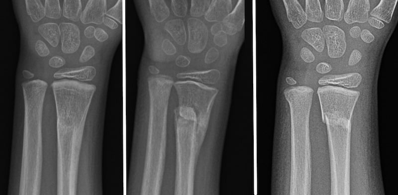
Bones that are fractured near the growth plate have immense potential for remodeling, which means reshaping themselves into a healthy alignment without surgical intervention.
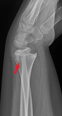
Examples of wrist fracture
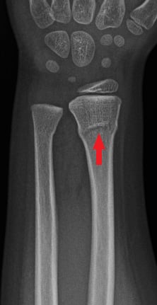
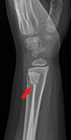
X-rays of the left wrist. Image A shows the front view (anteroposterior); image B shows the side view (lateral). The red arrow shows a fracture in the arm bone near the wrist (distal radius fracture). There is also a fracture near the wrist in the ulna, the other bone in the forearm.
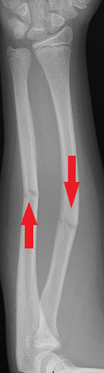
Example of remodeling process
Types of forearm fractures
Physeal fractures
These involve the growth plate in the wrist, typically of the radius. Because your child’s body can do an excellent job reshaping the fractured bones correctly, the fracture alignment has a wide range of acceptable positions, either with or without reduction (repositioning the bone without surgery) and subsequent casting. Your provider will also monitor for prolonged growth disturbance with x-rays, which typically occurs around 6 months from the time of the injury.
Examples of physeal fractures
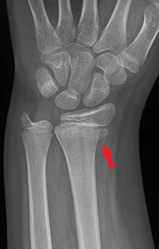
X-rays of the left wrist. Image A shows the front view (anteroposterior); image B shows the side view (lateral). The red arrows show a Salter-Harris II fracture of the forearm near the wrist (distal radius). A Salter-Harris Type II fracture is a fracture that goes through the thicker part of a long bone and the growth plate—a layer of tissue near the ends of a child’s bone—but doesn’t affect the end of the bone.
Torus (buckle) fractures
The most common forearm fracture, where one side of the bone is compressed but the other side remains strong. Depending upon the age and activity level of your child, these can be treated with a removable wrist brace or a cast.
Example of a torus fracture
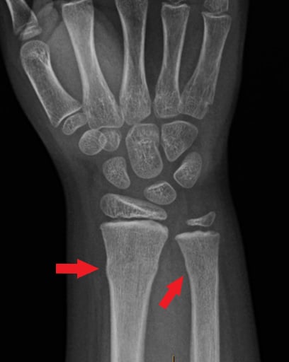

X-rays of the right wrist. Image A shows the front view (anteroposterior); image B shows the side view (lateral). The left arrows on each image show a buckle fracture of the forearm bone (radius) and the right arrows show a buckle fracture of the other forearm bone (ulna). A buckle fracture is when the bone bends but does not break all the way across.
Forearm shaft fractures
Although there is good potential for remodeling (reshaping), particularly in younger children, your provider may recommend additional treatment to correct the fracture so your child still has normal forearm rotation (palm up and palm down). This can frequently be achieved with closed reduction and casting, but does require surgery at times. There is also a prolonged risk for repeat fracture for several months, so your child will typically be transitioned to a removable brace and counseled on timing and specifics for return to full activities.
Examples of forearm shaft fractures
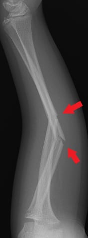
X-ray of the left forearm. Image A shows the front view (anteroposterior); image B shows the side view (lateral). The right arrows on each image show a fracture of the radius shaft and the left arrows show a fracture of the ulna shaft.