Burns: What's the Diagnosis?
Visual Diagnosis - March 2023
Column Author: Phil Jurasinski, DO | Internal Medicine-Pediatrics Resident, PGY 2
Column Editor: Joe Julian, MD, MPHTM | Hospitalist, Internal Medicine-Pediatrics; Clinical Associate Professor, Internal Medicine & Pediatrics
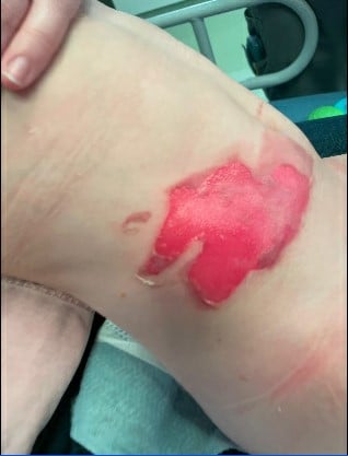
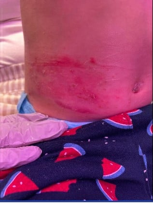
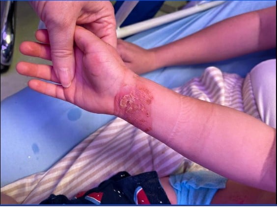
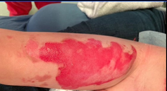
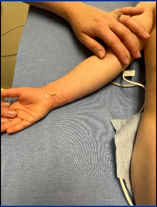
A 3-year-old male presents to the emergency department (ED) for evaluation of a burn. The patient and his mother were at a coffee shop that morning when the table that a hot cup of coffee was sitting on accidentally tipped over. The hot cup of coffee fell over onto the patient causing scald burns to his right wrist, right lower abdomen and right anterior thigh. Mother immediately removed his clothes, saw the burns, and brought him straight to the ED for further evaluation. He sustained no other injuries. Prior to this incident, mom reports that the patient was in his normal state of health with no recent illnesses, systemic symptoms or exposure to sick contacts. Mom states that the patient is otherwise healthy and his immunizations (including tetanus) are up to date.
Examination
Vital Signs: Temperature: 37.1 C, HR 118, RR 34, SpO2 99%, Pain 10/10
General: Alert, crying but consolable, in no acute respiratory distress.
HEENT: No burns on face, clear rhinorrhea with crying.
Respiratory: Non-labored respirations, lungs clear to auscultation bilaterally.
Cardiovascular: Tachycardic, regular rhythm, pulses equal in all extremities, normal peripheral perfusion with a capillary refill <2 seconds.
Gastrointestinal: Soft and non-distended, non-tender on non-burn areas, normal bowel sounds.
GU: Normal male genitalia for age. Circumcised phallus. Testes descended bilaterally. No scrotal edema or erythema. No burns.
MSK: Moving all extremities spontaneously and equally. Normal muscle bulk and tone. No deformities.
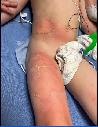
Integumentary: Warm and well perfused. Burns to the right volar wrist, right lower abdomen and right anterior thigh. Estimated total body surface area (TBSA) of 3.75%.
Neurologic: No focal deficits.
Pictures of the involved areas are shown below.
Question #1
How would these burns be classified?
- Superficial thickness
- Superficial partial thickness
- Deep partial thickness
- Full thickness
Answer #1
B. Superficial partial thickness burn
These burns are classified as superficial partial thickness burns, as they blanch with pressure, weep, are painful and will develop blisters. They may appear superficial at first, but can later appear as previously described within 12-24 hours. These wounds typically heal within seven to 21 days; necrotic debris may appear on the surface, making them likely to experience bacterial colonization. Superficial burns remain dry and do not form blisters, but will blanch with pressure and turn red. By day three, the pain and erythema will subside, and by day four, new epidermis will form. Deep partial thickness burns will be painful only to pressure, are wet or waxy dry, involve hair follicles and glandular tissue, and heal in approximately two to nine weeks. Full thickness burns involve all layers of the dermis and often injure subcutaneous tissue. They can be waxy white to leathery gray, can be painful or non-painful, and hair follicles can be easily removed.1
Question #2
How should this burn be treated?
- Mild oral analgesic and no dressing
- Wash with soap and water, apply topical ointment, change dressings daily
- Use an extended dressing for five to 10 days
- Use of a meshing instrument
Answer #2
C. Use an extended dressing for five to 10 days
Most clinicians now use extended dressings, which do not require daily wound care, and for larger wounds, do not require hospitalization. Loose blisters can be gently debrided, but tight blisters should be allowed to remain. Smaller amounts of pain medication can also be used for treatment.2 Silver sulfadiazine can impair reepithelization of the wound, and bacitracin can cause a rash if used for more than one week. Mild oral analgesics are appropriate for superficial burns and do not require dressings; this superficial partial thickness burn will require more treatment. A wound that is open for more than two weeks may require skin grafting for treatment. Meshing instruments are recommended only for full thickness burns that require skin grafting.2
Non-thermal burns may require special treatment. Chemical burns should be treated with irrigation of saline for 20 minutes and avoiding neutralizing agents. Tar burns should be cooled with water and may require the use of polymyxin. Electrical burns should be treated by removing the source of the electricity, and may involve complications of myoglobinuria, rhabdomyolysis and arrhythmia although the entrance and exit wounds may be small.3
Clinical Course
For the superficial partial thickness scald burns to his right wrist, right lower abdomen and right thigh, the patient was given two doses of intranasal fentanyl for pain and received a 20 mL/kg NS fluid bolus. The burn team was consulted and recommended wound debridement with soap and water. They dressed it with Mepilex-AG and Hypafix tape, recommended acetaminophen and ibuprofen as needed for pain, and discharged the patient home. His burns were redressed on his first visit to the burn clinic. At a subsequent visit one month later, his burns had fully healed.
References:
- American Burn Association White Paper. Surgical management of the burn wound and use of skin substitutes. Copyright 2009. https://ameriburn.org/
- Greenhalgh DG. Management of burns. N Engl J Med. 2019;380(24):2349-2359.
- Waitzman AA, Neligan PC. How to manage burns in primary care. Can Fam Physician. 1993;39:2394-2400. PMID: 8268745. PMCID: PMC2379923.
See all the articles in this month's Link Newsletter
Stay up-to-date on the latest developments and innovations in pediatric care - read the March issue of The Link.