Injured Finger
Visual Diagnosis - October 2023
Column Author: Philip Jurasinki, DO | Resident PGY 3
A previously healthy 3-year-old male presents to Children Mercy’s Emergency Department (ED) due to finger injury. Parents state that patient’s right third fingertip was accidentally shut in a car door three days ago. The patient seemed to be getting better; however, today the fingertip seems more swollen to the parents. There is bruising noted under the nail, however no open lacerations. The patient can move his finger normally and denies pain. No other injuries noted.
ROS: Positive for ecchymosis and joint swelling; negative for all other systems.
T: 36.6 C HR: 106 bpm RR: 24 BR/min BP: 109/58 SPO2: 99% Pain Score: 0
Exam: Right third finger with notable bruising/likely resolving subungual hematoma, with intact nailbed and CRT <2 seconds. Sensation intact; able to flex and extend digit at DIP. No nailbed laceration. Otherwise normal exam.
What is the next best step in management of the subungual hematoma?
- Trephinate (open up nail bed), do not perform radiography
- Do not trephinate, perform radiography
- Remove nail bed, do not perform radiography
- Remove nail bed, perform radiography
Answer: B
Trephination is recommended when hematomas are less than 48 hours old, are painful, and are associated with intact nail folds. Trephination can be done with an electrocautery, insulin syringe needle for aspiration, or boring needle with a double bevel 23 or single bevel 18 gauge needle.1-3 Beyond this point, trephination is not recommended, since most hematomas have clotted at this point, and the procedure would not relieve symptoms.1,3 Removal of the nail bed should typically occur when there is a nail laceration that threatens the preservation of the nail or digit. If a subungual hematoma involves over 50% of the nail bed, then radiography should be performed to assess whether there is a fracture.4 Distal phalanx fractures with subungual hematomas are considered open fractures, and need further management by a hand surgeon.1,4
Here is the radiograph that was obtained:
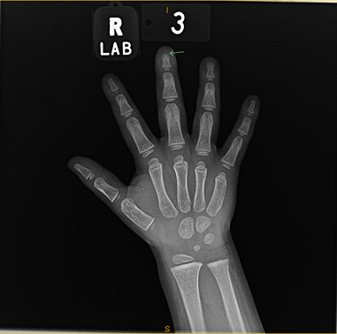
What is the diagnosis?
- Seymour fracture
- Shaft fracture
- Tuft fracture
- Mallet finger
- Jersey finger
This patient has a simple tuft fracture, as seen by the chip of bone at the end of the phalanx. Tuft fractures are the most common form of fracture associated with a subungual hematoma; they only need to be splinted. Seymour fractures are Salter-Harris type I or II fractures or juxta-epiphyseal fractures. They require evaluation by a hand surgeon, and potentially antibiotics. The shaft is intact on the radiograph, and the patient has no point tenderness over the shaft of the finger, ruling out a shaft fracture. Mallet fingers are caused by forced flexion of the extended DIP joint, resulting in avulsion of the extensor tendon. Jersey fingers are forced extension at the flexed DIP joint. They are often seen in football players, and present with bruising or swelling at the proximal stump. Mallet and jersey fingers are ruled out since he has normal range of motion at the DIP joint, and since the mechanism of injury does not match the radiograph.5,6
Case Follow-up:
The ED staff had communicated and verified the findings of the X-ray with orthopedics, who recommended splinting, and no follow-up. The patient’s clotted hematoma eventually resorbed, and he was instructed to follow up with his pediatrician. After eight weeks of splinting, the fracture had healed.
References:
- Patel L. Management of simple nail bed lacerations and subungual hematomas in the emergency department. Pediatr Emerg Care. 2014;30(10):742-745.
- Kaya TI, Tursen U, Baz K, Ikizoglu G. Extra‐fine insulin syringe needle: an excellent instrument for the evacuation of subungual hematoma. Dermatol Surg. 2003;29(11):1141-1143.
- Bonisteel PS. Trephining subungual hematomas. Can Fam Physician. 2008;54(5):693-693.
- Roser SE, Gellman H. Comparison of nail bed repair versus nail trephination for subungual hematomas in children. J Hand Surg. 2008;24(6):1166-1170.
- Bernstein ML, Chung KC. Hand fractures and their management: an international view. Injury. 2006;37(11):1043-1048. PMID: 17045997. doi:10.1016/j.injury.2006.07.020
- Cheah AE, Yao J. Hand fractures: indications, the tried and true and new innovations. J Hand Surg. 2016;41(6):712-722.
See all the articles in this month's Link Newsletter
Stay up-to-date on the latest developments and innovations in pediatric care – read the September issue of The Link.