What's the Diagnosis?
December 2021
Visual Diagnosis

Column Editor: Joe Julian, MD, MPHTM, FAAP | Hospitalist, Internal Medicine - Pediatrics | Clinical Associate Professor, Internal Medicine and Pediatrics, UMKC School of Medicine
A 4-month-old male is seen in the emergency department for further evaluation and management of a red and swollen left eye. Mother states that he has always had drainage from his left eye related to a clogged tear duct, but the swelling and redness are new. There is no known trauma. Associated symptoms include cough and nasal congestion but no fevers. He was taken to his primary care physician the previous day due to worsening swelling and redness; purulent nasolacrimal drainage was noted along with acute otitis media. Oral amoxicillin-clavulanate and topical erythromycin were prescribed. About 90 minutes after administration of both medications, the swelling around the eye worsened. Due to progressive swelling, the patient was brought to the emergency department for further evaluation.
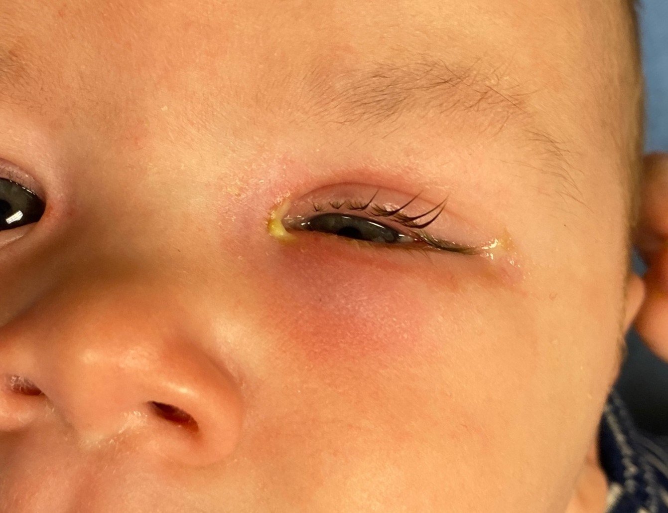
Vital signs are within age-appropriate ranges. Examination is notable for edema and erythema of the left inferior eyelid with purulent drainage medially (see image). The left conjunctiva has mild erythema. Pupils are round and reactive bilaterally and there are no deficits in eye movements noted. There is no proptosis. The child is non-toxic in appearance.
Which of the following is the most likely diagnosis?
A. Acute dacryocystitis
B. Orbital cellulitis
C. Periorbital hemangioma
D. Periorbital dermatitis
Answer: A. Acute dacryocystitis
The most likely diagnosis in this patient is acute dacryocystitis, an acute infection of the lacrimal sac and its surrounding tissues. Dacryocystitis typically occurs in the setting of congenital nasolacrimal duct obstruction (CNLDO) where both proximal and distal drainage of the lacrimal sac are impaired (most commonly caused by a dacryocystocele). Factors thought to contribute to acute dacryocystitis include anatomical (narrow drainage system) and immune-related (immature immune system) causes. The patient has a history of chronic eye drainage consistent with CNLDO. The picture shows significant inferomedial edema with erythema spreading to the periorbital tissues and purulent drainage is present medially from the punctum (exit of canaliculi from lacrimal sac). These indicate an acute infection and a diagnosis of acute dacryocystitis.
Orbital cellulitis is a complication of acute dacryocystitis. The patient’s examination does not support this diagnosis. Patient findings that point away from orbital cellulitis include intact eye movements, no proptosis, no pupillary defect, lack of severe edema of the eyelid and globe, and lack of systemic findings such as fever. If there is concern for any orbital involvement, a CT scan with contrast can be obtained to evaluate for such.
Periorbital hemangioma does not fit given the acuity of the presentation and the lack of prior dermatologic findings. Red/purple nodules would be present in the congenital type of hemangioma, while mobile red flat/nodular lesions would be present in the infantile type. These are not described historically or seen on examination, and are not consistent with the patient’s longstanding eye drainage.
Periorbital dermatitis should be on the differential given the perceived worsening after administration of the prescribed medications (both oral and topical). Allergic etiologies are usually bilateral, but the unilateral topical application makes single eye involvement a possibility. However, this involvement would not explain the patient’s initial erythema, edema and purulent drainage.
Which of the following treatments is the most appropriate next step?
A. Ampicillin-clavulanate IV
B. Topical erythromycin
C. Lacrimal duct probing
D. Ceftriaxone + clindamycin IV
Answer: A. Ampicillin-clavulanate IV
Regardless of any intervention, antibiotics are the most appropriate next step. The most common pathogens for pediatric acute dacryocystitis are Staphylococcus aureus, Streptococcus pneumoniae, coagulase-negative Staphylococcus, and alpha-hemolytic Streptococcus. Of note, these data are obtained from limited studies/series of pediatric patients in the post-vaccine era and differ significantly from those in the adult population. Since community-acquired methicillin-resistant Staphylococcus aureus (MRSA) is uncommon in this condition, coverage in a patient who is not systemically ill is probably not necessary. A beta-lactam plus a beta-lactamase inhibitor (e.g., ampicillin-sulbactam, amoxicillin-clavulanate) is a reasonable choice that allows for simple transition to an oral regimen. Ceftriaxone + clindamycin IV is unnecessarily broad for this patient but would be appropriate for one that is systemically ill-appearing.
Even if lacrimal duct probing was considered in this patient, intravenous antibiotic administration would be necessary prior to any intervention given the risk of probing-induced bacteremia. However, the frequency of this risk is under some debate. Conservative management with antibiotic administration is an appropriate next step while evaluating for surgical intervention. Additionally, literature evaluating the efficacy of probing is limited. Topical antibiotics do not have much of a role in acute infection and are typically used in cases of chronic dacryocystitis.
Case Conclusion:
CT imaging was obtained in the emergency department to evaluate for complications such as preorbital/orbital cellulitis. Results were notable for a dacryocystocele, a dilated lacrimal duct and thickening/inflammation consistent with dacryocystitis. The patient was given a single dose of ampicillin IV and admitted. This treatment was expanded to ampicillin-sulbactam IV. Ophthalmology evaluated the patient and recommended adding moxifloxacin eyedrops with outpatient follow-up. The patient improved on ampicillin-sulbactam and was transitioned back to amoxicillin-clavulanate to complete 10 total days of antibiotic therapy. Of note, parainfluenza was detected on the patient’s respiratory pathogen panel, which likely explains the nasal congestion/cough and could have been a contributing factor to his presentation.
Aerobic/anaerobic cultures of eye drainage yielded polymicrobial growth: Streptococcus anginosus, Chryseobacterium gleum, coagulase-negative Staphylococcus, diphtheroids, and two Prevotella species. While some of these pathogens may not necessarily be covered by amoxicillin-clavulanate, the patient improved clinically, and his symptoms were nearly resolved by his ophthalmology follow-up two days after discharge. For this reason, no changes were made to his antimicrobial regimen.
References:
- Ali MJ. Pediatric acute dacryocystitis. Ophthalmic Plast Reconstr Surg. 2015;31(5):341-347.
- Goodyear PWA, Firth AL, Strachan DR, Dudley M. Periorbital swelling: the important distinction between allergy and infection. Emerg Med J. 2004;21(2):240-242.
- Kuchar A, Lukas J, Steinkogler FJ. Bacteriology and antibiotic therapy in congenital nasolacrimal duct obstruction. Acta Ophthalmol Scand. 2000;78(6):694-698.
- Williams KJ, Allen RC. Paediatric orbital and periorbital infections. Curr Opin Ophthalmol. 2019;30(5):349-355.