What’s the Diagnosis? A New Facial Nodule
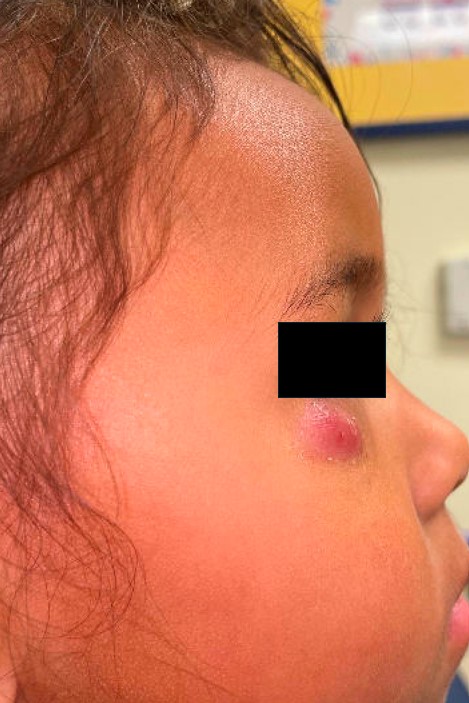
A 3-year-old girl presented with a solitary lesion below her right eye that had been present for approximately three months.
The lesion initially appeared as a small, skin-colored bump that was thought to be a stye, but it slowly enlarged over time. During the past several weeks, it developed a purple-red discoloration. The lesion was described as mildly tender and occasionally pruritic, but there was no associated fever or systemic symptoms.
Her parents noted no bleeding, though a small amount of tan drainage was observed the day before presentation, which resolved spontaneously. The family had been applying topical metronidazole cream for about one week with minimal improvement.
There was no relevant family history, no chronic medical conditions, and no history of recurrent styes or ocular disease.
Physical examination
On the right upper cheek, inferior to the right lower eyelid, there was a 1 × 2 cm dark red–violaceous firm nodule and mild peripheral scale. The lesion was nonfluctuant and nontender with no overlying warmth, ulceration, or crusting. The “teeter-totter” sign was negative. There was no evidence of periorificial dermatitis, or other periocular lesions.

Question 1:
What is the most likely diagnosis?
A. Idiopathic facial aseptic granuloma (IFAG)
B. Epidermal inclusion cyst
C. Chalazion
D. Pilomatricoma
E. Pyogenic abscess
Correct answer: A. Idiopathic facial aseptic granuloma (IFAG)
Idiopathic facial aseptic granuloma (IFAG) is a benign pediatric granulomatous dermatosis classically seen in infants and young children. It most commonly presents as a solitary, firm, red to violaceous papule or nodule on the cheek or periocular area. Lesions commonly enlarge slowly over weeks to months and are typically painless or only mildly tender; systemic symptoms are usually absent. Many lesions resolve spontaneously over several months to about a year, though individual courses vary. These clinical features align with this patient’s age, location of the lesion, slow enlargement, violaceous color change, and absence of systemic signs.
The exact cause of IFAG remains unknown. Early infectious hypotheses have not been supported by consistent microbiologic evidence: special stains and cultures are typically negative. Multiple retrospective series and reviews have described clinical and histologic overlap between IFAG and childhood granulomatous rosacea or periorificial dermatitis, prompting the view that IFAG may lie on a spectrum with these entities. Shared features include granulomatous perifollicular inflammation, periocular/cheek distribution, and reported responses in some cases to treatments commonly used for rosacea (topical metronidazole or ivermectin; systemic macrolide antibiotics or doxycycline.)
This patient’s age, lesion location and color, slow progression, nonfluctuant firm texture, ultrasound impression compatible with IFAG, and lack of systemic signs strongly support IFAG as the leading diagnosis. The brief episode of tan drainage could reflect minor surface breakdown but is not definitive for bacterial abscess; cultures and special stains are often negative in IFAG. The presence of prior drainage/punctum supports close observation and reassessment rather than reflexive incision and drainage.
Epidermal inclusion cyst:
Epidermal inclusion cysts are firm, mobile, dome-shaped nodules with a central punctum and are filled with keratinous debris. They are typically skin-colored or yellow-white rather than violaceous, and they can emit a foul-smelling keratinous material if ruptured. They are uncommon in young children and rare on the eyelids. Ultrasound often shows a well-circumscribed hypoechoic lesion with internal echoes consistent with keratin rather than the homogeneous solid echotexture of IFAG.
Chalazion:
A chalazion results from chronic lipogranulomatous inflammation of a blocked meibomian gland within the tarsal plate. Clinically, it presents as a subcutaneous eyelid nodule that is often palpable through the tarsus and may have mild overlying erythema. It tends to be intrapalpebral rather than subcutaneous on the cheek or infraorbital skin. Some cohorts report prior or concurrent chalazia in a subset of patients with IFAG. Careful attention to exact anatomic location and eyelid/meibomian gland anatomy can help distinguish the two.
Pilomatricoma:
Pilomatricoma is a common benign adnexal neoplasm in children. They are usually firm to hard, skin-colored or bluish, and often show the “teeter-totter sign” on palpation due to calcification where pressing on one end of the nodule causes the other to raise up in response. Lesions are frequently found on the face or upper extremities of children but tend to be hard rather than compressible or tender. Ultrasound typically shows a well-defined lesion with internal echogenic foci of calcification, absent in IFAG. Histologically, pilomatricoma shows basaloid cells and shadow (“ghost”) cells, distinct from the granulomatous infiltrate in IFAG.
Pyogenic abscess:
A bacterial abscess is acutely inflamed, warm, tender, and fluctuant, often with overlying erythema, drainage of purulent material, and systemic findings such as fever or regional lymphadenopathy. Ultrasound reveals a central hypoechoic or anechoic cavity with posterior acoustic enhancement representing pus. In contrast, IFAG is typically afebrile, firm, nonfluctuant, and demonstrates a solid hypoechoic lesion without fluid collection. Misdiagnosis of IFAGs as abscesses can result in unnecessary scarring in cosmetically sensitive areas.
Question 2:
What is the best next step in management?
A. Immediate surgical excision and biopsy
B. Incision and drainage with oral antibiotics
C. Conservative management with observation and follow-up
D. Oral clarithromycin for 8 weeks
E. Oral prednisone taper
Correct answer: C. Conservative management with observation and follow-up
Observation with close clinical follow-up is the preferred initial approach for an otherwise well child with a lesion clinically consistent with IFAG because many lesions resolve spontaneously and invasive interventions (incision and drainage, excision) carry cosmetic risk. Families should be counseled about the usual benign course and advised to return if the lesion enlarges rapidly, becomes increasingly tender/warm, drains frankly purulent material, or if systemic symptoms develop.
Although high-quality randomized data are lacking, observational case series and reports describe several medical options in persistent, progressive, symptomatic, or cosmetically concerning lesions:
• Topical agents used for rosacea (metronidazole, topical ivermectin) have been used and may be trialed for limited lesions.
• Oral antibiotics (clarithromycin, erythromycin or doxycycline) have been described as effective in some series and case reports but supporting studies are limited. Treatment courses are typically several weeks. Antibiotic therapy may be reasonable when families request active management, when lesions are multiple, or when rapid improvement is desired, but the uncertain benefit should be discussed alongside stewardship considerations.
• Intralesional corticosteroid injection (e.g., triamcinolone) has been used for persistent nodules to reduce size and inflammation and may be considered when cosmesis is a priority and the lesion is accessible; systemic corticosteroids are not routinely recommended. Intralesional injections may not be well tolerated especially in younger children and carry risks such as atrophy and dyspigmentation, which could be very cosmetically impactful on the face.
• Excision or biopsy is reserved for diagnostic uncertainty, atypical features (rapid growth, ulceration, recurrence after treatment), or failure of conservative/medical therapy. This is because of the risk of scarring and impact on cosmesis given the cosmetically prominent location of many IFAGs. In addition recurrence after biopsy or excision is common. Unfortunately IFAGs are frequently misdiagnosed as abscesses resulting in unnecessary and ineffective incision and drainage resulting in a scar but without significant improvements in the lesion.
In short: observation is first-line; consider topical or systemic therapies for multiple, or cosmetically significant lesions after shared decision-making.
Key points and clinical pearls:
- Idiopathic facial aseptic granuloma (IFAG) typically appears as a solitary, firm, red to violaceous papule or nodule on the cheek or periocular area of young children.
• Etiology is unclear; microbiology is usually negative, and clinical/histologic overlap with granulomatous rosacea or periorificial dermatitis has been reported.
• Histology shows granulomatous perifolliculitis with multinucleated giant cells; special stains/cultures usually fail to identify organisms.
• Lesions may be solitary or multiple. Periocular IFAG can mimic chalazion.
• Ultrasound often demonstrates a solid, hypoechoic lesion without a central fluid collection.
• Observation and reassurance are first-line; topical or systemic agents) or intralesional corticosteroid are options for persistent, multiple, or cosmetically significant lesions. Avoid routine incision and drainage as this is not typically helpful and likely to result in scarring.
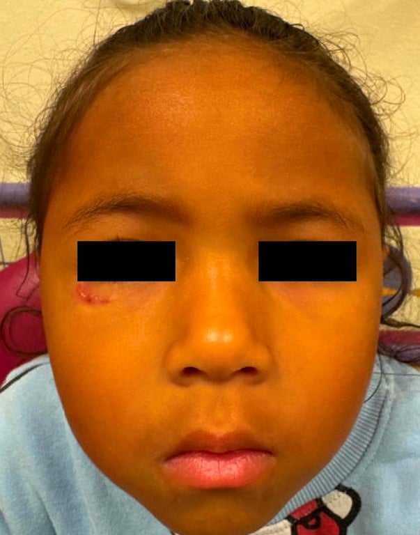
Case update:
4 months after patient’s initial visit the lesion had decreased significantly in size and appeared to be involuting.
References:
- Wortsman X, et al. Idiopathic facial aseptic granuloma: updated review of an evolving clinical entity. Actas Dermosifiliogr (Engl Ed). 2019.
- Mendes SR, et al. Idiopathic facial aseptic granuloma. BMJ Case Rep. 2022.
- Weir SA, et al. Idiopathic facial aseptic granuloma: case series and review. Pediatr Dermatol. 2023.
- Sánchez-Espino LF, et al. Idiopathic facial aseptic granuloma: report of successful treatment. JAAD Case Rep. 2022.
Associate Program Director, Pediatric Dermatology Fellowship; Assistant Professor of Pediatrics, University of Missouri-Kansas City School of Medicine; Clinical Assistant Professor of Internal Medicine, University of Kansas School of Medicine