Visual Diagnosis: Acquired Lighter Patches
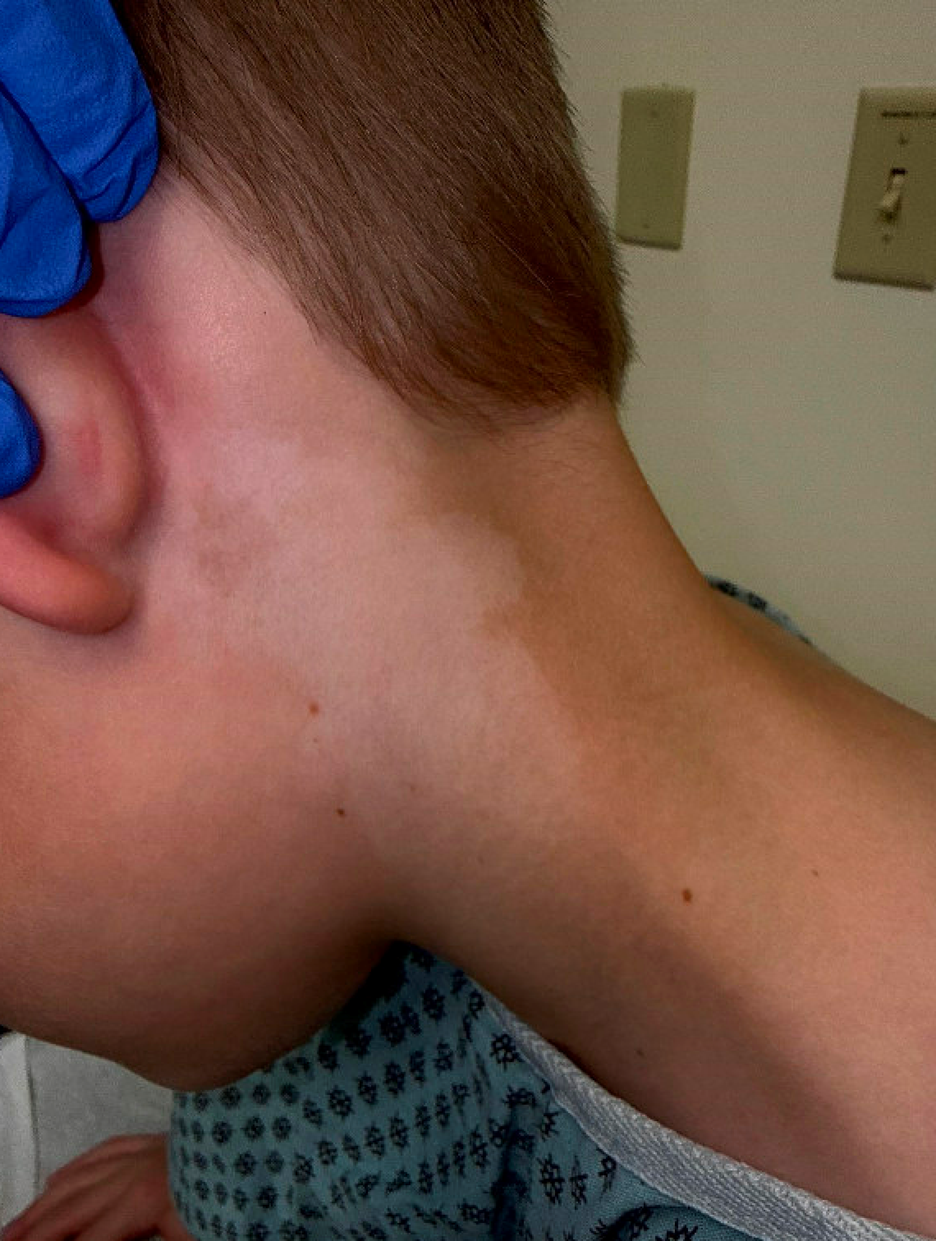
A 6-year-old boy presents for evaluation of light-colored patches on the skin. Approximately one year ago, his parents noticed a lighter patch on the left side of his neck as well as a similar area behind his knee. The lesion behind the knee has since resolved, but the patch on the left neck has persisted. The lesion has remained stable in size since it was first noticed.
The family is not applying any topical agents to the area. Family history is notable for the patient’s father having similar lighter patches on his skin, but he has not received a diagnosis. There are no associated symptoms, including pruritus or pain, and the family denies a preceding rash.
The patient is otherwise healthy and growing well. Review of systems is negative for fatigue, weight changes, temperature intolerance, headaches, gastrointestinal symptoms, or changes in sleep or energy.
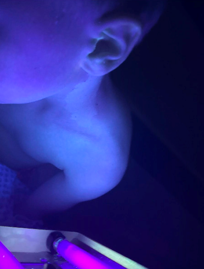
Examination, including examination under Wood’s lamp, reveals the findings below:
Question 1
Which of the following is the most likely diagnosis?
- Pityriasis alba
B. Nevus depigmentosus
C. Post-inflammatory hypopigmentation
D. Tinea versicolor
E. Vitiligo
Correct Answer: E
Vitiligo
Explanation:
On physical examination, there is a well-demarcated depigmented patch on the left lateral neck without scale or textural change. Examination under Wood’s lamp reveals bright blue-white fluorescence with sharp borders corresponding to the lesion. These findings are in keeping with acquired areas of depigmentation consistent with vitiligo. Vitiligo is an acquired disorder characterized by complete depigmentation resulting from autoimmune destruction of melanocytes. Lesions are typically well demarcated and accentuate under Wood’s lamp examination, showing bright blue-white fluorescence with sharp borders. When it occurs on hair-bearing skin, it may be associated with depigmentation of the hair, known as poliosis. Vitiligo has a variable and unpredictable natural history, particularly in children. Some patients experience slow progression or stability, while others may develop additional lesions over time. Spontaneous repigmentation can occur, especially in children and in sun-exposed areas, but is often incomplete. Early intervention may improve cosmetic outcomes and promote repigmentation, particularly in localized disease. Importantly, vitiligo is a benign, noncontagious condition and does not affect overall physical health, though it may have significant psychosocial impact.
- Pityriasis alba presents as hypopigmented (not depigmented) patches, often in children with atopic dermatitis, xerosis cutis or seborrheic dermatitis. Lesions have ill-defined borders, may show subtle scale and do not demonstrate bright fluorescence under Wood’s lamp.
- Nevus depigmentosus is usually congenital or presents in early infancy and remains stable in size and distribution over time. “Nevus depigmentosus” is a misnomer, as lesions are usually hypopigmented, not depigmented. Wood’s lamp examination does not show the characteristic enhancement seen in vitiligo.
- Post-inflammatory hypopigmentation follows an inflammatory dermatosis such as eczema, infection or trauma. A history of a preceding rash is typically present, borders are less distinct and repigmentation usually occurs gradually over time. Wood’s lamp findings are subtle or absent.
- Tinea versicolor commonly affects the trunk and proximal extremities and is associated with fine scale. Lesions are usually hypopigmented, not depigmented. Under Wood’s lamp, lesions may fluoresce yellow-green, and diagnosis can be confirmed with potassium hydroxide examination.
Question 2
Which of the following is the most appropriate initial treatment for this patient’s condition?
- Observation only, as treatment is ineffective
B. Oral corticosteroids
C. High-potency topical corticosteroids applied daily for several months
D. Topical calcineurin inhibitor applied to affected areas daily
E. Topical ruxolitinib cream applied to affected areas daily
Correct Answer: D
Topical calcineurin inhibitor applied to affected areas
Explanation:
For localized vitiligo in children, particularly on the face and neck, topical calcineurin inhibitors such as tacrolimus or pimecrolimus are first-line therapy. These agents promote repigmentation while avoiding the risk of skin atrophy associated with prolonged topical corticosteroid use in sensitive areas.
Observation alone may be reasonable in select cases, but early treatment may improve cosmetic outcomes. Oral corticosteroids are not indicated for localized, stable disease. High-potency topical corticosteroids may be used for limited durations on non-facial areas but are not preferred on the neck in young children.
Topical ruxolitinib, a Janus kinase inhibitor, is approved by the Food and Drug Administration for the treatment of nonsegmental vitiligo in patients 12 years of age and older. Although it represents an important therapeutic advance, it is not approved for use in this patient due to age and would not be appropriate as initial therapy in a 6-year-old child.
Question 3
Which of the following statements regarding vitiligo and thyroid disease in children is most accurate?
- All children with vitiligo should undergo routine thyroid laboratory screening at diagnosis.
B. Thyroid disease occurs only in children with segmental vitiligo.
C. Children with vitiligo have an increased risk of autoimmune thyroid disease, but routine screening is not required in asymptomatic patients.
D. Thyroid disease is rare in pediatric vitiligo and screening is unnecessary even if symptoms develop.
E. Thyroid dysfunction typically precedes the onset of vitiligo in most children.
Correct Answer: C
Children with vitiligo have an increased risk of autoimmune thyroid disease, but routine screening is not required in asymptomatic patients.
Explanation:
Children with vitiligo have a higher prevalence of autoimmune thyroid disease compared with the general pediatric population. However, most guidelines do not recommend routine thyroid screening in asymptomatic children at the time of vitiligo diagnosis. Instead, thyroid testing should be targeted to patients with suggestive symptoms, a strong family history, extensive or progressive disease or other autoimmune conditions. Pediatricians should counsel families about this association and maintain a low threshold for evaluation if symptoms arise.
References:
- Ezzedine K, Eleftheriadou V, Whitton M, van Geel N. Vitiligo. Lancet. 2015;386(9988):74-84.
- Silverberg JI, Silverberg NB. Association between vitiligo and thyroid disease in children. Pediatr Dermatol. 2015;32(5):e216-e221.
- Kakourou T, Kanaka-Gantenbein C, Papadopoulou A, Kaloumenou E, Chrousos GP. Increased prevalence of chronic autoimmune thyroiditis in children and adolescents with vitiligo. J Am Acad Dermatol. 2005;53(2):220-223.
- Mazereeuw-Hautier J, Brenaut E, Dubois D, et al. Thyroid abnormalities in children with vitiligo: A prospective study. Br J Dermatol. 2012;167(4):892-896.
- Taïeb A, Picardo M. Clinical practice. Vitiligo. N Engl J Med. 2009;360(2):160-169.
- Rosmarin D, Pandya AG, Lebwohl M, et al. Ruxolitinib cream for the treatment of vitiligo: two randomized controlled trials. N Engl J Med. 2022;387(16):1445-1455.
- American Academy of Dermatology Association. Guidelines of care for the management of vitiligo. J Am Acad Dermatol. 2023.
Associate Program Director, Pediatric Dermatology Fellowship; Assistant Professor of Pediatrics, University of Missouri-Kansas City School of Medicine; Clinical Assistant Professor of Internal Medicine, University of Kansas School of Medicine