Evidence Based Strategies: Pediatric Scoliosis: An Evidence-Based Framework for Early Identification and Management
Adolescent idiopathic scoliosis is a common condition affecting approximately 1%–3% of adolescents, most of whom never require treatment.1 However, severe cases may result in restrictive pulmonary disease, chronic pain and, rarely, cardiopulmonary compromise. The condition is characterized by a three-dimensional spinal deformity with lateral curvature measuring at least 10 degrees in addition to vertebral rotation. Idiopathic scoliosis is classified by age at diagnosis (infantile, juvenile or adolescent). Non-idiopathic forms of scoliosis are classified by etiology (e.g., neuromuscular, syndromic, congenital). In infantile and juvenile idiopathic scoliosis, severe deformity raises particular concern for impaired pulmonary development and long-term cardiopulmonary function. In contrast, adolescent idiopathic scoliosis more commonly presents concerns related to pain and psychosocial well-being.
How to diagnose scoliosis
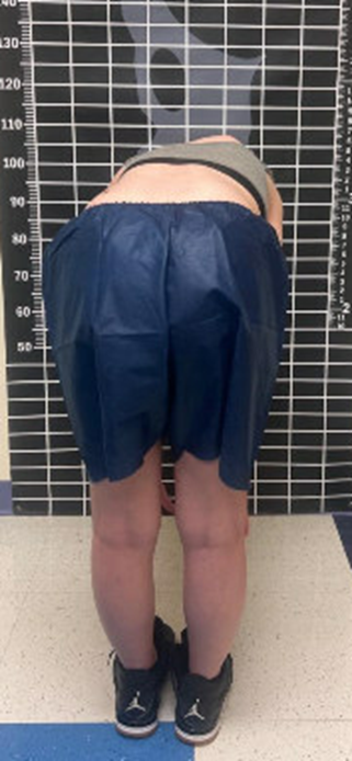
Most cases of scoliosis are diagnosed based on physical examination followed by radiographs. The Adams forward bend test (Figure) and quantification of trunk rotation using a scoliometer are widely accepted screening tools for scoliosis. Rotation >5° should prompt the screening clinician to obtain upright radiographs.2 In younger children or those with medical comorbidities that prevent participation in the examination, the presence of rib prominence, flank prominence, pelvic obliquity or shoulder tilt should also prompt radiographic imaging. If scoliosis is confirmed radiographically (Cobb angle >10°), a comprehensive history and physical examination should be performed to evaluate for secondary causes of scoliosis, with special attention to the neurologic examination and family history.
Routine magnetic resonance imaging (MRI) is not indicated for all patients with scoliosis. However, those with abnormal neurologic examination, early onset scoliosis (age <10 years), congenital scoliosis or certain syndromic conditions such as neurofibromatosis and Marfan syndrome are more likely to have intraspinal pathology. Therefore, they are more likely to benefit from MRI evaluation.3
When to refer to a spine specialist
Referral to a pediatric spine specialist is generally recommended for patients with Cobb angles ≥15°, with brace treatment initiated between 20° and 25°, depending on the patient’s age and curve characteristics. Spinal bracing has been shown to be effective but imperfect in arresting the progression of the curve in growing children. Emerging evidence suggests that modern three-dimensional bracing systems may offer improved curve control compared with earlier brace designs.4,5 Therefore, early identification and timely referral to initiate bracing may reduce the likelihood of surgical intervention, especially in younger patients at higher risk of curve progression.
Surgical intervention is generally considered when coronal curves exceed 50°, as curves beyond this threshold are highly likely to progress despite skeletal maturity, although smaller curves may progress as well.6 The mainstay of treatment is posterior spinal fusion, but in younger patients, there are a variety of growth-friendly surgical strategies to preserve spinal and thoracic growth.
Where can I find more information?
Children’s Mercy provides a clinical pathway available at Scoliosis. The Scoliosis Clinical Pathway provides decision support and guidance to both primary care and specialty clinic practitioners for the screening, evaluation and management of scoliosis.
References:
- Li M, Nie Q, Liu J, Jiang Z. Prevalence of scoliosis in children and adolescents: a systematic review and meta-analysis. Front Pediatr. 2024;12:1399049. doi:10.3389/fped.2024.1399049
- Labelle H, Richards SB, De Kleuver M, et al. Screening for adolescent idiopathic scoliosis: an information statement by the Scoliosis Research Society International Task Force. 2013;8:17. doi:10.1186/1748-7161-8-17
- Li AW, Chang A, Murphy JS, et al. Current practices in MRI screening in early onset scoliosis. Spine Deform. 2025;13(3):961-966. doi:10.1007/s43390-024-01033-4
- Weinstein SL, Dolan LA, Wright JG, Dobbs MB. Effects of bracing in adolescents with idiopathic scoliosis. N Engl J Med. 2013;369(16):1512-1521. doi:10.1056/NEJMoa1307337
- Weiss HR, Turnbull D, Seibel S, Kleban A. First end-result of a prospective cohort with AIS treated with a CAD Chêneau style brace. J Phys Ther Sci. 2019;31(12):983-991. doi:10.1589/jpts.31.983
- Weinstein SL, Ponseti IV. Curve progression in idiopathic scoliosis. J Bone Joint Surg Am. 1983;65(4):447-455.
Figure. A 14-year-old female with thoracic scoliosis demonstrating the Adams forward bend test. Note the asymmetric rib prominence, with an elevated right posterior rib cage.
Clinical Assistant Professor of Orthopedic Surgery, University of Missouri-Kansas City School of Medicine; Research Assistant Professor of Orthopedic Surgery, University of Kansas School of Medicine