
Visual Diagnosis: What’s the Diagnosis? Cutaneous Mastocytoma
Case Presentation
A 13-month-old boy presents for evaluation of a slowly enlarging lesion on the upper back. His parents first noticed a small red spot at approximately 5 months of age that they initially thought was a mosquito bite.
Rather than resolving, the lesion gradually enlarged and developed intermittent episodes of increased redness and occasional blistering. During these episodes, the lesion also appears to spread slightly before returning to its original size and light brown color.
The lesion does not appear painful or pruritic, and no clear trigger has been identified. There are no similar lesions elsewhere on his body. His parents deny episodes of flushing, shortness of breath, vomiting, diarrhea or fainting.
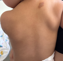
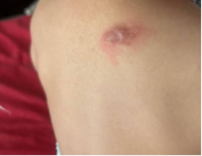
On physical exam: approximately 2 × 1 cm light-brown thin plaque with irregular borders on the upper back (left). Gentle rubbing of the lesion leads to erythema and swelling of and surrounding the lesion, as shown in the photo provided by parents (right).
Question 1: What is the most likely diagnosis?
- Café-au-lait macule
- Congenital melanocytic nevus
- Solitary cutaneous mastocytoma
- Arthropod bite reaction
- Recurrent bullous impetigo
Correct Answer: (C) Solitary cutaneous mastocytoma
Solitary cutaneous mastocytoma is a form of cutaneous mastocytosis that usually presents in infancy or early childhood as a solitary yellow-brown or reddish-brown papule, plaque or nodule.1,2 Lesions can intermittently become erythematous, swollen or blister because of mast-cell mediator release.
Several features in this case favor a mastocytoma. The lesion appeared during infancy, persisted beyond the expected duration for an insect bite and slowly enlarged. Most importantly, the lesion displays a positive Darier sign: after physical irritation to the area, the lesion and surrounding skin become erythematous and edematous, and blisters develop.
Incorrect Answers:
A café-au-lait macule would typically remain flat and uniformly pigmented without episodic erythema or blistering (A). A congenital melanocytic nevus would not be expected to repeatedly urticate (B). An arthropod bite should resolve rather than persist and enlarge over many months (D). Recurrent bullous impetigo would be expected to produce more acute superficial vesicles or bullae rather than a persistent brown plaque between episodes (E).
Question 2: Which is an essential element of initial management for this patient?
- Surgical excision of the lesion
- Systemic corticosteroids until the plaque resolves
- Avoidance of specific medications
- Prophylactic oral antibiotics
Correct Answer: (C) Avoidance of specific medications
Families should receive individualized counseling about factors that may provoke mast-cell mediator release, including medications such as polymyxin B, aspirin, alcohol, dextromethorphan and certain anesthetic agents.3
Incorrect Answers:
Most childhood solitary mastocytomas follow a benign course and tend to improve or involute over time, so do not require surgical excision.1-3 Treatment is therefore directed primarily at symptoms and prevention of mast-cell degranulation rather than removal of the lesion (A).
Topical medications may be beneficial. For this patient, the family was instructed to apply triamcinolone 0.1% ointment twice daily during episodes of increased redness until the inflammation resolves. An antihistamine can be considered if the lesion becomes significantly pruritic or swollen or episodes are frequent; however, neither systemic corticosteroids nor oral antibiotics are necessary, and the lesion itself will not resolve with their use (B and D).
Question 3: Several months later, the parents call because the child has developed recurrent flushing and chronic diarrhea. Which laboratory test would be most useful as part of the initial evaluation?
- Serum total IgE
- Serum tryptase
- Antinuclear antibody
- Blood culture
Correct Answer: (B) Serum tryptase
Most children with a solitary mastocytoma have disease limited to the skin and do not require an extensive systemic evaluation. The absence of flushing, gastrointestinal symptoms, syncope, generalized urticaria or organomegaly in this patient at presentation was therefore reassuring so no further workup was indicated at that time.
If symptoms suggestive of more extensive mast-cell mediator release develop, however, basal serum tryptase can be used as a surrogate marker of mast-cell burden and followed longitudinally.2,3
An elevated tryptase level in a pediatric patient must be interpreted differently than in an adult. Children with cutaneous mastocytosis may have elevated serum tryptase without systemic mastocytosis, so an elevated value by itself does not automatically mandate bone marrow biopsy.2,3 Findings such as persistent systemic symptoms, organomegaly, extensive skin disease or other clinical abnormalities should guide the need for additional evaluation.
Parents should therefore be instructed to report new persistent flushing, chronic diarrhea, fainting, generalized hives or blistering/new lesions.
Mastocytoma Tips for Pediatricians
- Consider mastocytoma when an infant or young child presents with a persistent yellow, red or brown plaque that periodically becomes red, swollen or blistered.
- Look for the Darier sign: urtication or erythema after gentle rubbing of the lesion; however, a negative Darier sign does not exclude the diagnosis.
- Avoid unnecessary invasive testing in an otherwise well child with a typical solitary lesion. Systemic mastocytosis is rare in children with isolated cutaneous disease.
- Determine if further evaluation may be warranted. Ask about systemic symptoms including recurrent flushing, chronic diarrhea, syncope, generalized urticaria or extensive/new blistering.
- Serum tryptase may be useful if symptoms are concerning, but an elevated tryptase in a child with cutaneous mastocytosis does not always indicate systemic disease.
- Management is generally conservative and includes reassurance, symptom control and individualized counseling regarding avoidance of mast-cell triggers.
- The prognosis is favorable. Most skin lesions that erupt by 2 years of age spontaneously resolve before puberty.
References:
- Leung AKC, Lam JM, Leong KF. Childhood solitary cutaneous mastocytoma: clinical manifestations, diagnosis, evaluation, and management. Curr Pediatr Rev. 2019;15(1):42-46. doi:10.2174/1573396315666181120163952
- Ługowska-Umer H, Czarny J, Rydz A, Nowicki RJ, Lange M. Current challenges in the diagnosis of pediatric cutaneous mastocytosis. Diagnostics (Basel). 2023;13(23):3583. Published December 1, 2023. doi:10.3390/diagnostics13233583
- Schaffer JV. Pediatric mastocytosis: recognition and management. Am J Clin Dermatol. 2021;22(2):205-220. doi:10.1007/s40257-020-00581-5