Visual Diagnosis: An 8-Year-Old Boy with Hair Loss - Case Presentation
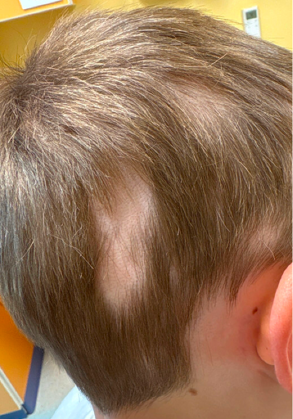
An 8-year-old boy with a history of atopic dermatitis presents for evaluation of hair loss. His parents first noticed a small bald patch on the back of his scalp approximately two months ago. Since then, a second patch has appeared on the left side of his scalp. The areas are asymptomatic, and he denies pain, itching or increased hair shedding. There has been no recent illness, fever, hospitalization, weight loss or medication changes.
On examination, there are two well-circumscribed patches of hair loss on the right and left occipital scalp. The underlying skin appears smooth without scale, erythema or scarring. The follicular ostia are present. Several short hairs tapering toward the scalp are noted at the margins of the patches. There is no cervical or occipital lymphadenopathy. There is regular pitting of the nails. The eyebrows and eyelashes are normal.
Question 1
Which of the following is the most likely diagnosis?
A. Alopecia areata
B. Tinea capitis
C. Trichotillomania
D. Telogen effluvium
E. Traction alopecia
Answer
- Alopecia areata
Discussion
Alopecia areata is an autoimmune disorder characterized by non-scarring hair loss. Children typically present with one or more smooth, well-demarcated patches of hair loss with normal-appearing scalp skin. Exclamation point hairs (short hairs that taper proximally near the scalp surface) are a classic finding and suggest active disease. The follicular ostia, or follicular openings, are usually preserved and still visible in alopecia areata, unlike scarring types of hair loss. Regular nail pitting can be seen in alopecia areata and may indicate a less favorable prognosis.
When evaluating a child with hair loss, careful examination of the scalp can often distinguish alopecia areata from other common causes without additional testing.
- Tinea capitis is the most common cause of patchy hair loss in prepubertal children and is frequently mistaken for alopecia areata. Unlike alopecia areata, the scalp is usually abnormal, with findings such as scale, erythema, black dots, broken hairs, crusting, pustules or even a boggy inflammatory plaque in the case of a kerion. Occipital or posterior cervical lymphadenopathy is a particularly helpful clue that favors tinea capitis over alopecia areata. Children may also report scalp pruritus or tenderness. A dermatophyte culture of the scale from the lesion can confirm the diagnosis.
- Trichotillomania results from repetitive hair pulling and typically produces irregularly shaped patches rather than the round or oval patches seen in alopecia areata. Examination often reveals hairs of varying lengths due to repeated breakage and regrowth, with incomplete hair loss within affected areas. The scalp skin is usually normal, although excoriations, erosion or bleeding may occasionally be present. A history of stress, anxiety or other repetitive behaviors (onychophagia, skin picking, lip licking, etc.) may be present, although many children and families are unaware of the hair-pulling behavior.
- Telogen effluvium causes diffuse hair shedding rather than discrete patches of alopecia. It usually develops several months after a physiologic or emotional stressor, such as a significant illness, surgery, hospitalization, rapid weight loss or major psychosocial stress. Families often report excessive hair accumulation on pillows, brushes or in the shower. Examination reveals generalized decreased hair density with a normal scalp rather than focal bald patches.
- Traction alopecia results from chronic tension on the hair shaft from hairstyles such as tight braids, ponytails, buns, extensions or cornrows. Hair loss most commonly affects the frontal and temporal scalp and follows the distribution of tension. Early disease may show broken hairs and retained hairs along the frontal hairline, known as the “fringe sign.” Unlike alopecia areata, a careful hairstyle history usually reveals the underlying cause.
This patient’s smooth, well-circumscribed patches of hair loss, normal scalp skin and exclamation point hairs are most consistent with alopecia areata.
Clinical Pearl: When evaluating a child with hair loss, first determine whether the hair loss is patchy or diffuse, whether the scalp appears normal or abnormal, and whether the hairs are missing entirely or broken at different lengths. These observations will correctly identify the cause of hair loss in many children.
Question 2
Which of the following is the most appropriate laboratory evaluation for this patient?
A. CBC, comprehensive metabolic panel, iron studies, ANA and thyroid function tests.
B. Thyroid function testing if there are symptoms, examination findings or risk factors suggesting thyroid disease.
C. Routine autoimmune screening in all children with alopecia areata.
D. Food allergy testing.
E. Extensive endocrine evaluation.
Answer
b.Thyroid function testing only if there are symptoms, examination findings or risk factors suggesting thyroid disease.
Discussion
The diagnosis of alopecia areata is clinical, and routine laboratory testing is not recommended for most children with localized disease.
Targeted testing may be considered when there are symptoms, examination findings or risk factors suggesting an associated condition. Thyroid disease is the most commonly discussed autoimmune association. Thyroid screening may be appropriate in children with clinical symptoms of thyroid dysfunction, Down syndrome, a personal history of autoimmune disease or a strong family history of thyroid disease.
Although alopecia areata is associated with atopic conditions, including atopic dermatitis, allergic rhinitis and asthma, atopic dermatitis alone does not require broad laboratory screening. Food allergy testing is not indicated for alopecia areata.
Clinical Pearl: Most children with alopecia areata do not require laboratory testing. Reserve thyroid screening and other investigations for children with suggestive symptoms, examination findings or significant risk factors.
Question 3
What is the most appropriate initial management for this patient?
A. Begin high-potency topical corticosteroid therapy and monitor for regrowth.
B. Prescribe topical ruxolitinib cream.
C. Start oral ritlecitinib therapy.
D. Reassure the family that treatment is unnecessary because spontaneous regrowth always occurs.
E. Begin oral biotin supplementation.
Answer
- Begin high-potency topical corticosteroid therapy and monitor for regrowth.
Discussion
High-potency topical corticosteroids are considered first-line therapy for children with limited patch-type alopecia areata. Commonly used agents include clobetasol 0.05% solution, foam or ointment applied to affected scalp areas. Treatment is generally continued for several months while monitoring for regrowth and local adverse effects such as skin atrophy or folliculitis.
Many children with localized disease experience spontaneous regrowth, but the course is unpredictable and recurrence is common. Because treatment is relatively safe and may accelerate regrowth, many families choose to pursue therapy.
Topical ruxolitinib is approved by the Food and Drug Administration (FDA) for atopic dermatitis and nonsegmental vitiligo but is not FDA-approved for alopecia areata, and current evidence is insufficient to support routine use. Oral ritlecitinib is FDA-approved for severe alopecia areata in patients 12 years of age and older and is generally reserved for more extensive disease. Biotin supplementation is not helpful unless true biotin deficiency is present.
Additional treatment options may include intralesional corticosteroids in older cooperative children and adolescents, topical minoxidil as an adjunctive therapy, oral minoxidil and topical irritants.
For children with acute, rapidly progressive alopecia areata, systemic corticosteroids may occasionally be used to try to halt disease progression or serve as a bridge to other therapies; however, relapse after discontinuation is common.
Clinical Pearl: For a child with one or two patches of alopecia areata, high-potency topical corticosteroids remain the recommended first-line treatment and can often be initiated while awaiting dermatology evaluation.
Summary:
Alopecia areata presents with smooth, well-demarcated patches of non-scarring hair loss. The absence of scale helps distinguish it from tinea capitis, while hairs of varying lengths suggest trichotillomania. Routine laboratory screening is unnecessary for most children, and high-potency topical corticosteroids remain first-line therapy for localized disease.
References:
- Xu L, Liu KX, Senna MM. A practical approach to the diagnosis and management of hair loss in children and adolescents. Front Med (Lausanne). 2017;4:112.
- Patel D, Li P, Bauer AJ, Castelo-Soccio L. Screening guidelines for thyroid function in children with alopecia areata. JAMA Dermatol. 2017;153(12):1307-1310.
- Barton VR, Toussi A, Awasthi S, Kiuru M. Treatment of pediatric alopecia areata: a systematic review. J Am Acad Dermatol. 2022;86(6):1318-1334.
Associate Program Director, Pediatric Dermatology Fellowship; Assistant Professor of Pediatrics, University of Missouri-Kansas City School of Medicine; Clinical Assistant Professor of Internal Medicine, University of Kansas School of Medicine
Director, Quality & Safety; Director, Outpatient Antimicrobial Stewardship Program; Division Director, Infectious Diseases; Medical Director, Vaccines for Children (VFC) Program; Professor of Pediatrics, University of Missouri-Kansas City School of Medicine; Clinical Assistant Professor of Pediatrics, University of Kansas School of Medicine