
Visual Diagnosis: A Chronic Pruritic Rash
A 13-year-old girl with Fitzpatrick type V skin and a lifelong history of a poorly controlled itchy rash presents for her first dermatology visit. Her mother reports persistent itching that worsens at night and interferes with sleep and school performance. She has been treated intermittently with over-the-counter moisturizers and low-, medium-, and high-potency topical corticosteroids.
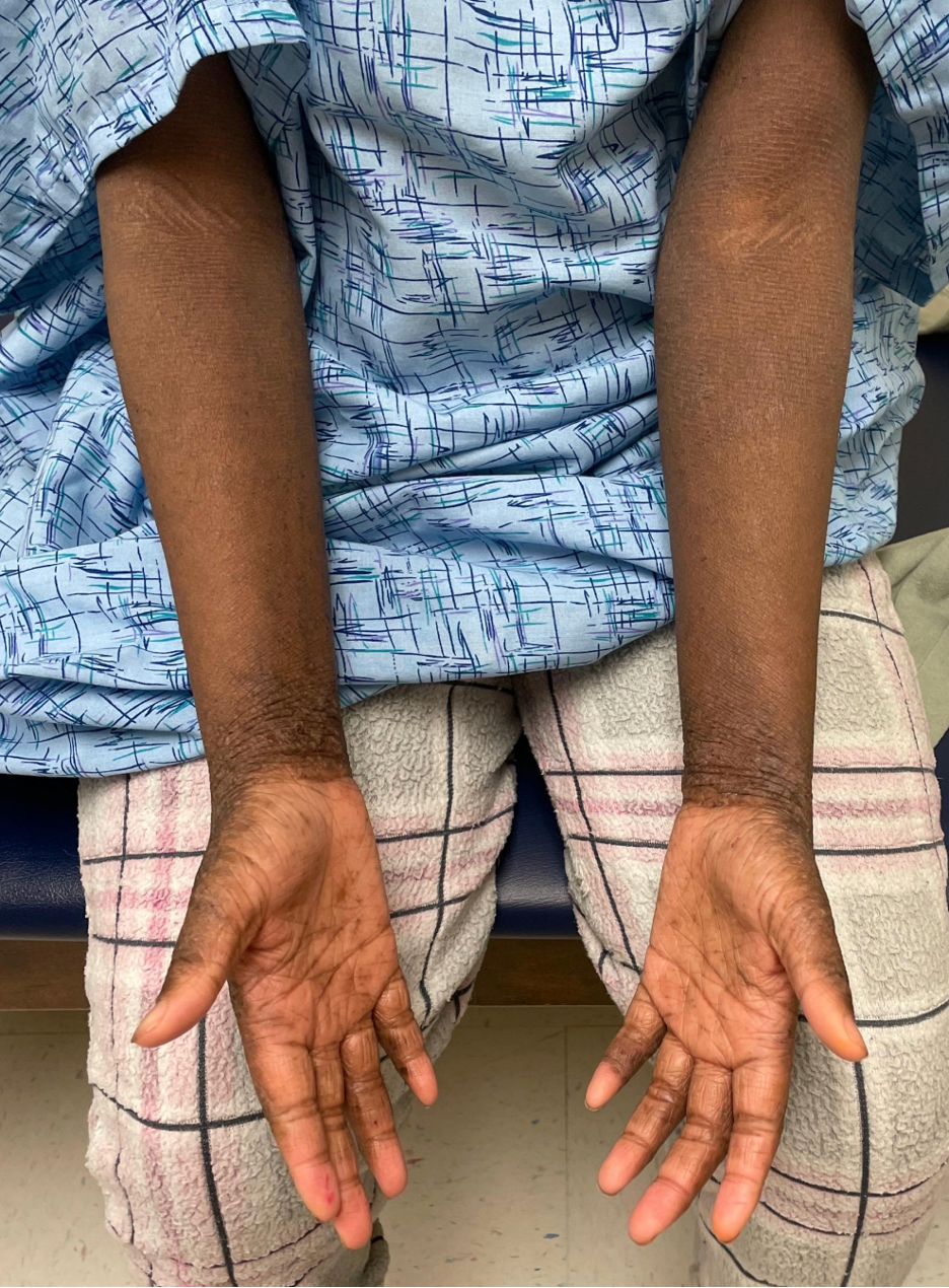
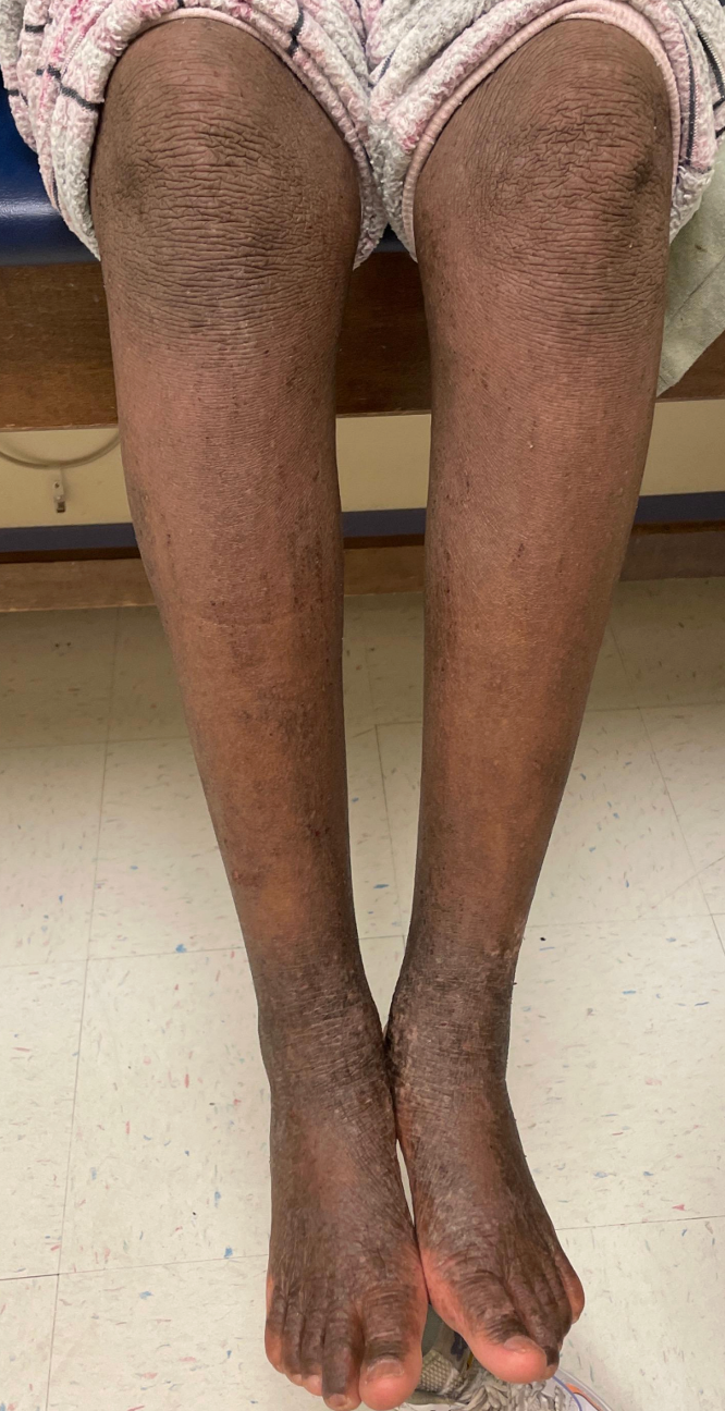
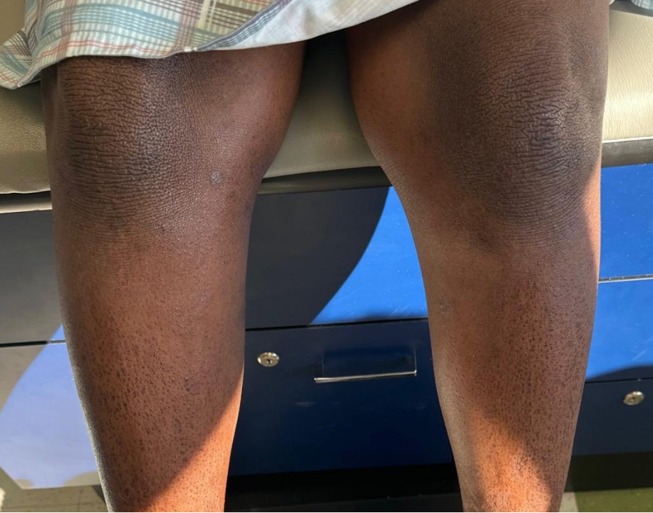
On examination, there are well-defined, thickened (lichenified), dark brown, slightly erythematous plaques on both wrists, the inner elbows and behind both knees. On closer inspection, the palms have exaggerated, deepened skin lines, and polygonal scales are present on both shins. There are no vesicles, pustules, honey-colored crusting, burrows or dome-shaped papules.
Several conditions can contribute to eczema flares or complicate atopic dermatitis in children.
Question 1:
Based on the clinical photographs, which of the following is most likely contributing to this patient’s presentation?
- Secondary bacterial infection (Staphylococcus aureus impetiginization)
- Eczema herpeticum
- Molluscum contagiosum with molluscum dermatitis
- Ichthyosis vulgaris
- Scabies infestation
Answer: D
The photographs demonstrate two classic findings of ichthyosis vulgaris (IV): hyperlinear palms and fine polygonal (“plate-like”) scaling on the shins. These features form the hallmark of IV, the most common inherited skin barrier disorder. IV is caused by loss-of-function mutations in the filaggrin gene (FLG), which encodes a protein essential for maintaining skin barrier integrity.
Recognizing IV in a child with AD is clinically important: FLG mutations are the strongest known genetic risk factor for AD and mark a more severe phenotype. AD patients with concurrent IV have higher rates of allergic rhinoconjunctivitis. IV is also linked to earlier onset of eczema and greater AD severity. This supports the “outside-inside” hypothesis, where a leaky skin barrier allows allergens to penetrate the skin, driving transcutaneous sensitization and the “atopic march” from eczema to food allergies, rhinitis, and asthma.
Each of the other answer choices represents a common complicator of AD in children that pediatricians should be able to recognize. Children with AD are at significantly increased risk for skin infections due to their compromised skin barrier, altered immune responses and disrupted skin microbiome. Superinfections are among the most common triggers of eczema flares and can cause substantial disease worsening.
- Secondary bacterial infection (S. aureus impetiginization): S. aureus colonizes the skin of most patients with AD, and true bacterial superinfection is the most common infectious complication. It presents as honey-colored (golden) crusting, weeping, oozing, and tenderness overlying eczematous plaques— not the dry, lichenified plaques seen in this patient. Impetiginization should be suspected when a previously stable area of eczema suddenly worsens, becomes painful or develops purulent drainage. Management focuses on treating the underlying eczema (which itself restores skin barrier defenses and reduces S. aureus colonization), with short-course anti-staphylococcal antibiotics reserved for cases with overt impetiginization. An aerobic culture can be helpful to confirm impetiginization and direct/narrow antibiotic therapy. Dilute bleach baths can be used as an adjunctive measure to reduce bacterial burden. Importantly, routine antimicrobial treatment is not recommended in AD patients without signs of overt infection.
- Eczema herpeticum: This is a potentially life-threatening disseminated herpes simplex virus (HSV) infection that occurs almost exclusively in patients with AD. It presents with a distinctive eruption of monomorphic, dome-shaped vesicles that rupture to form “punched-out” erosions with hemorrhagic crusting, typically clustered in areas of active eczema, most commonly the face, neck and trunk. Patients are often febrile and appear systemically unwell. The key clinical clue is the monomorphic nature of the vesicles/erosions; they generally appear similar in size and shape. This patient has no vesicles, punched-out erosions or hemorrhagic crusting. Eczema herpeticum requires prompt treatment with systemic antivirals (acyclovir or valacyclovir).
- Molluscum contagiosum with molluscum dermatitis: Molluscum contagiosum (MC) is a common poxvirus infection in children and is five times more prevalent in children with AD than in those without eczema. It presents as discrete, dome-shaped, skin-colored to pearly papules with a characteristic central dimple (umbilication). In children with AD, MC can trigger an eczematous reaction around the molluscum papules, known as “molluscum dermatitis,” which can be mistaken for an eczema flare. MC tends to be more widespread in AD patients due to autoinoculation via scratching. This patient has no dome-shaped umbilicated papules, making MC unlikely.
- Scabies infestation: Scabies is an important mimic of AD in children and should always be considered in the differential of a pruritic child, especially when eczema is “atypical” or treatment-resistant. It presents with intensely pruritic papules, excoriations and characteristic burrow: thin, wavy, gray-white lines most often found in the interdigital web spaces, flexural wrists, ankles and, in adolescents, the genital area. Nocturnal pruritus is prominent (as it is in AD), which can cause diagnostic confusion. The key distinguishing features are the burrows and the involvement of interdigital spaces and feet, areas typically spared in AD. While this patient has wrist involvement, there are no burrows, interdigital lesions or papules suggestive of scabies.
Question 2:
Given this patient’s presentation, which of the following is the most appropriate next step in management?
- Prescribe high-potency topical corticosteroids to all affected areas and reassess in four weeks.
- Initiate dupilumab.
- Start oral prednisone taper to achieve rapid disease control.
- Prescribe crisaborole ointment to all affected areas.
Answer: B
This patient has moderate-to-severe AD with widespread thickened plaques across multiple body sites, reflecting years of uncontrolled disease. The 2026 American Academy of Dermatology (AAD) Pediatric AD Guidelines and the 2024 American Academy of Allergy, Asthma and Immunology/American College of Allergy, Asthma and Immunology (AAAAI/ACAAI) Joint Task Force Guidelines both recommend that when optimal topical management is not enough to control eczema — or when a child has widespread, severe disease that significantly impairs quality of life — systemic therapy should be considered.
The AAAAI/ACAAI Joint Task Force strongly recommends dupilumab (with high-certainty evidence) for patients aged 6 months and older with moderate-to-severe AD who have not responded adequately to, are intolerant of, or are unable to use mid-potency or greater topical therapy. While this patient has not yet been managed by a dermatologist, her long history of poorly controlled disease and the degree of lichenification make it likely she will need systemic treatment. A brief trial of optimized topical therapy (to document inadequate response) is reasonable before starting dupilumab, but unnecessary delays in escalation should be avoided given her disease burden.
Why the other answers are wrong:
- Choice A (high-potency topical steroids everywhere): While effective short-term, prolonged use of high-potency topical steroids over large body areas raises concerns about skin thinning, stretch marks and systemic absorption — and would not address the overall disease burden. The question stem also notes the patient has already trialed high-potency topical steroids.
- Choice C (oral prednisone): The AAD strongly recommends against long-term systemic corticosteroids for pediatric AD due to growth suppression, adrenal insufficiency and rebound flaring upon discontinuation.
- Choice D (crisaborole monotherapy): Crisaborole is approved for mild-to-moderate AD and has modest efficacy. It is insufficient as sole therapy for disease of this severity.
Question 3:
Dupilumab is initiated for this patient. Which of the following best describes important counseling regarding this medication?
- Dupilumab is an immunosuppressant and increases the risk of serious infections; routine laboratory monitoring is required.
- Conjunctivitis is the most characteristic and most common ocular side effect, occurring in approximately 10% of adolescent patients.
- Dupilumab has been associated with an increased risk of malignancy in long-term pediatric studies.
- Dupilumab has no effect on atopic comorbidities such as asthma.
Answer: B
Per the U.S. Food and Drug Administration (FDA) label, the most common adverse reactions in AD (incidence ≥1%) are injection site reactions, conjunctivitis, blepharitis, oral herpes, keratitis, eye pruritus, other HSV infection, dry eye and eosinophilia, with injection site reactions and conjunctivitis being the most frequent. Injection site reactions are generally at least as common as conjunctivitis, but they are typically mild, transient and common with many subcutaneous biologic medications.
Conjunctivitis is the most characteristic and clinically important adverse effect of dupilumab, because it is specifically associated with the drug in AD (it does not appear to occur at elevated rates in the asthma, nasal polyp or eosinophilic esophagitis trials). In the adolescent AD trial, conjunctivitis occurred in about 10% of dupilumab-treated patients versus ~5% on placebo. In the trial of children aged 6 months to <6 years, all cases were mild and resolved. Higher baseline AD severity and a prior history of conjunctivitis increase the risk.
The mechanism is not fully understood but may relate to decreased mucin production by conjunctival goblet cells when IL-4 and IL-13 signaling is blocked.
How to manage conjunctivitis in practice:
- Mild cases (redness, dryness, tearing): Lubricating eye drops (artificial tears) and warm compresses are usually sufficient.
- Moderate cases (persistent symptoms): May require topical anti-inflammatory eye drops (corticosteroid or cyclosporine drops), ideally in consultation with ophthalmology.
- When to refer urgently: Blurred vision, photophobia, purulent discharge or eye pain warrant urgent ophthalmology evaluation.
- Discontinuation due to eye symptoms is rare (only about 5% of patients in some real-world studies).
Why the other answers are wrong:
- Choice A (immunosuppressant): Dupilumab is not particularly immunosuppressing. Unlike older systemic agents (methotrexate, cyclosporine, azathioprine), dupilumab selectively blocks IL-4 and IL-13, two cytokines that drive allergic inflammation. It does not broadly suppress the immune system and does not increase overall infection risk. In fact, skin infections tend to decrease on dupilumab, likely because the skin barrier improves. Routine blood work monitoring is not required, which is a significant practical advantage for pediatric patients.
- Choice C (malignancy risk): There is no signal for increased malignancy risk with dupilumab in pediatric or adult studies.
- Choice D (no effect on comorbidities): Emerging data suggest dupilumab may have benefits beyond the skin (see below).
Dupilumab is FDA-approved for moderate-to-severe AD in patients aged 6 months and older. It is administered as a subcutaneous injection every two to four weeks, with weight-based dosing in children. Beyond controlling eczema, several observational studies suggest broader benefits in children; it may slow the “atopic march” and may protect linear growth.
References:
- Guttman-Yassky E, Renert-Yuval Y, Brunner PM. Atopic dermatitis. Lancet. 2025;405(10478):583-596. doi:10.1016/S0140-6736(24)02519-4
- Davis DMR, Alikhan A, Bercovitch L, et al. Guidelines of care for the management of atopic dermatitis in pediatric patients. J Am Acad Dermatol. 2026;95(1):121.e1-121.e26. doi:10.1016/j.jaad.2026.02.113
- Schoch JJ, Anderson KR, Jones AE, Tollefson MM; Section on Dermatology. Atopic dermatitis: update on skin-directed management: clinical report. Pediatrics. 2025;155(6):e2025071812. doi:10.1542/peds.2025-071812.
- AAAAI/ACAAI JTF Atopic Dermatitis Guideline Panel, Chu DK, Schneider L, et al. Atopic dermatitis (eczema) guidelines: 2023 AAAAI/ACAAI Joint Task Force on Practice Parameters GRADE- and Institute of Medicine-based recommendations. Ann Allergy Asthma Immunol. 2024;132(3):274-312. doi:10.1016/j.anai.2023.11.009
- Paller AS, Simpson EL, Siegfried EC, et al. Dupilumab in children aged 6 months to younger than 6 years with uncontrolled atopic dermatitis: a randomised, double-blind, placebo-controlled, phase 3 trial. Lancet. 2022;400(10356):908-919. doi:10.1016/S0140-6736(22)01539-2