What's the Diagnosis?
January 2022
Visual Diagnosis

Column Editor: Joe Julian, MD, MPHTM, FAAP | Hospitalist, Internal Medicine - Pediatrics | Clinical Associate Professor, Internal Medicine and Pediatrics, UMKC School of Medicine
A previously healthy 2-month-old male is directly admitted for further evaluation of sustained tachypnea after daily evaluation by his PCP. Patient initially had acute otitis media, approximately four weeks ago, and received a course of amoxicillin which was completed. During his mother’s six-week postpartum checkup approximately two weeks ago, the midwife noted that the patient’s breathing was abnormal. Approximately one week ago, the patient developed more labored breathing and retractions. He was seen by his PCP, who ordered an echocardiogram and a chest X-ray which was notable for right-sided pneumonia. He has been taking amoxicillin for the past four days; azithromycin and albuterol were added two days ago without much improvement.
Patient was born at 39 weeks via spontaneous vaginal delivery and had an unremarkable nursery stay. Maternal labs including GBS, HIV, gonorrhea and chlamydia were negative per report. There is no family history of heart disease, pulmonary conditions or immunodeficiency. The patient has not had any associated fevers but has had a sporadic cough and a single episode of perioral cyanosis when upset recently. He has been feeding adequately but has gained no weight in three to four weeks.
- Vitals: Temp 37°C | Pulse 139 | Resp 56 | BP 116/61 | 97% on room air | Weight 5.1 kg
- General: comfortable, no acute distress
- Respiratory: mild tachypnea with subcostal retractions worsened in appearance due to pectus excavatum, significantly diminished aeration with dullness to percussion over the right lung fields, left lung fields with appropriate aeration
- Cardiovascular: regular rate and rhythm without murmur, normal S1/S2, pulses equal in all extremities
- Abdomen: soft and non-tender
- Extremities: warm and well-perfused, cap refill 2 seconds
Previous imaging from four days earlier not available but reports are as follows:
- Two-view chest X-ray shows right middle and lower lobe consolidation with right hemidiaphragm elevation due to loops of bowel
- Echocardiogram shows left to right shunting across PFO, right lower lobe pulmonary vein not seen but no anomalous return visualized (repeat study recommended)
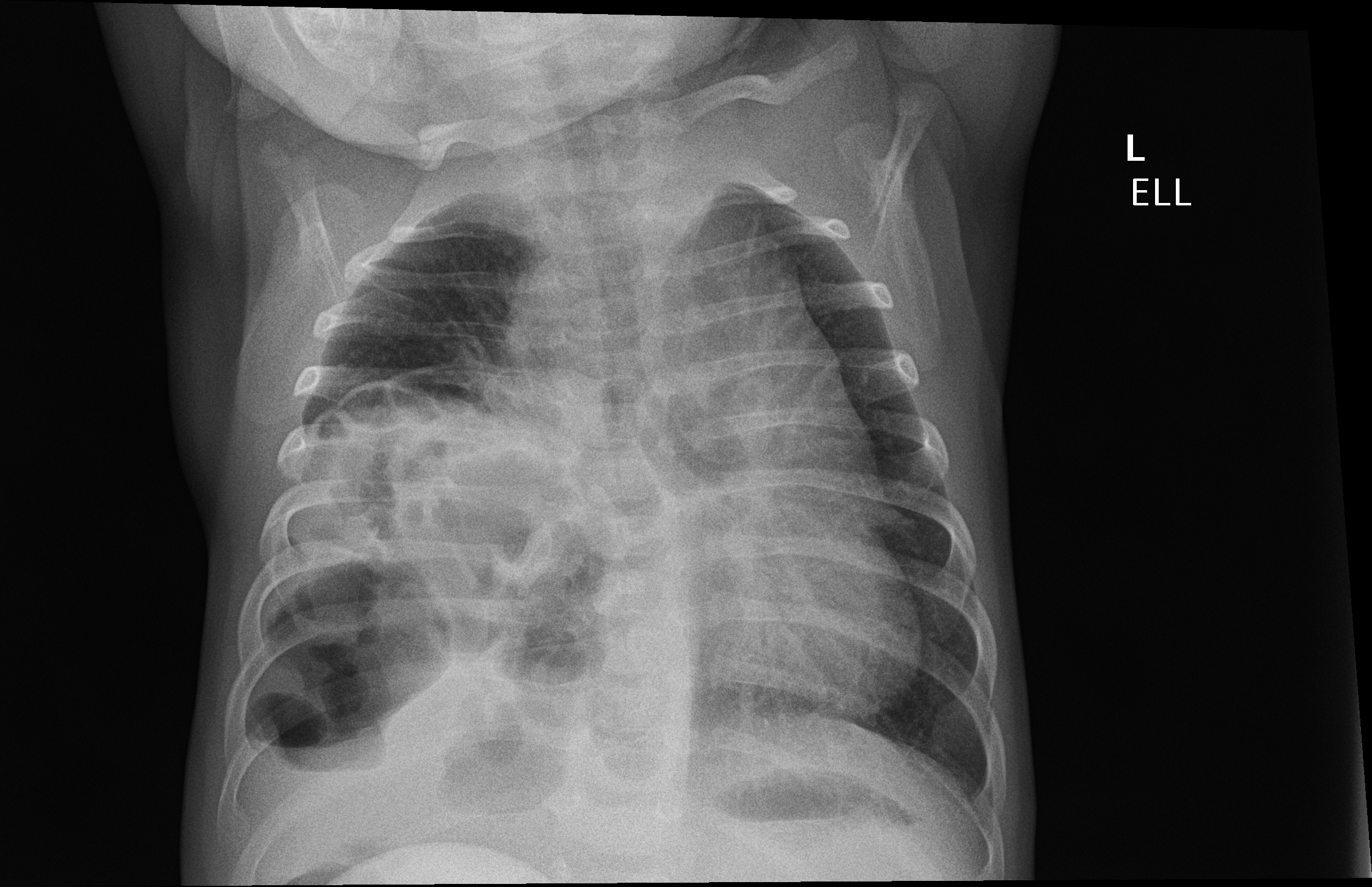
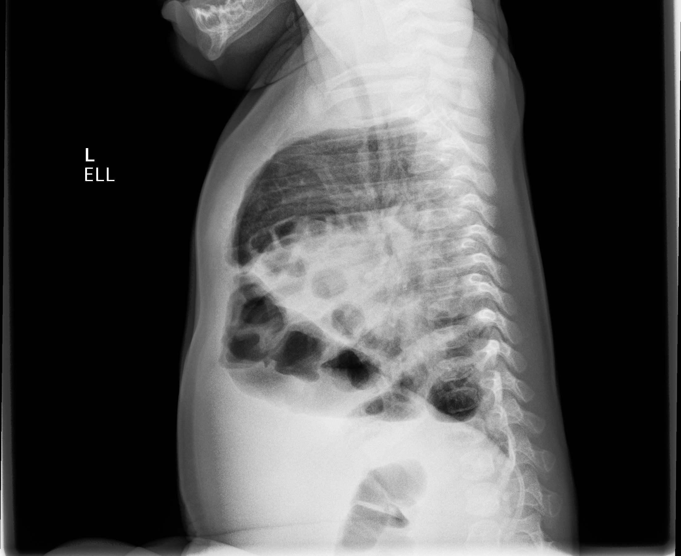
A two-view chest X-ray is obtained on admission and is shown here.
Which of the following is the most likely diagnosis?
A. Partial anomalous pulmonary venous return
B. Multifocal community-acquired pneumonia
C. Congenital diaphragmatic hernia
D. Congenital pulmonary airway malformation
Answer: C. Congenital diaphragmatic hernia
The chest radiograph demonstrates air-filled loops of bowel in the right lower chest with patchy atelectasis concerning for congenital diaphragmatic hernia. An additional differential diagnosis of marked diaphragmatic eventration (abnormal elevation of an intact diaphragm due to loss of muscle or innervation) was also considered.
This case demonstrates an atypical presentation of a congenital diaphragmatic hernia. The diagnosis is typically made in the prenatal or newborn period with respiratory distress and has significant risk of poor prognosis. Delayed presentations (defined as older than 30 days) tend to have significantly better outcomes and are perhaps “acquired,” with intestines possibly herniating through a defect previously occluded by spleen or liver. As with this patient, a common presenting symptom is failure to thrive. Right-sided hernias tend to present with respiratory symptoms (as opposed to either respiratory or gastrointestinal with left-sided ones). Misdiagnosis is common; pneumonia is the primary one. Misdiagnoses of pleural effusion or empyema tend to have greater morbidity, given the potential for visceral injury during intervention (such as thoracotomy or pleural drainage).
Partial anomalous pulmonary venous return is a condition where pulmonary veins enter the venous system, creating a left to right shunt. In hypogenetic lung syndrome (“scimitar syndrome”), the right pulmonary vein connects to the inferior vena cava. The syndrome is associated with hypoplastic right lung and pulmonary artery. While not all of the pulmonary veins were visualized on the echocardiogram, there was no anomalous return. The lack of visualization was likely due to anatomic displacement by the intestines. However, given the delayed presentation of a right-sided congenital diaphragmatic hernia, the initial echocardiogram findings should be re-evaluated due to the possibility of additional associated developmental defects.
Multifocal community-acquired pneumonia would not show the presence of air-filled bowel, as demonstrated in this patient’s radiograph. While not available for comparison, the previous radiograph may not have been as clear, and diaphragmatic eventration could have been a bigger concern. The patient has received adequate treatment with amoxicillin and the addition of azithromycin. If there is concern for a multifocal bacterial pneumonia that is not improving on standard beta-lactam therapy, Staphylococcus aureus must be considered as a pathogen (which is unlikely given the clinical history and scenario).
Congenital pulmonary airway malformation (previously known as congenital cystic adenomatoid malformation) is an abnormal maturation and proliferation defect leading to dysplastic lung tissue. There are three types of this condition with cysts of varying sizes having variable prognoses. In some instances, the cysts can have an appearance similar to herniated bowel and are difficult to differentiate without visualization of the bowel gas pattern. The clinical presentation varies depending upon size and location, but respiratory distress, recurrent pneumonia and failure to thrive should prompt its consideration.
With reference to the case, surgery was consulted and the patient was taken to the operating room on hospital day 3. A majority of the small bowel and right colon were found in the chest, confirming a diagnosis of congenital diaphragmatic hernia. Fortunately, the lung did not appear to be underdeveloped. An anterolateral defect was found and repaired with mesh via an intrathoracic approach. The patient was discharged on hospital day 4 after an uneventful recovery. The patient was noted to have significant improvement in respiratory symptoms and weight gain at a four-week follow-up appointment.
References:
- Baerg J, Kanthimathinathan V, Gollin G. Late-presenting congenital diaphragmatic hernia: diagnostic pitfalls and outcome. Hernia. 2012;16:461-466.
- Congenital Diaphragmatic Hernia Study Group. Late-presenting congenital diaphragmatic hernia. J Pediatr Surg. 2005;40:1839-1843.
- Congenital heart disease. In: Wells RG, ed. Diagnostic Imaging of Infants and Children. McGraw Hill; 2013.
- Developmental abnormalities of the lungs and diaphragm. In: Wells RG, ed. Diagnostic Imaging of Infants and Children. McGraw Hill; 2013.
- Hon KL, Fung RCM, Leung AKC. Delayed presentation of congenital diaphragmatic hernia with acute respiratory distress: challenges in diagnosis and management. Case Rep Pediatr. 2020.