What's the Diagnosis?
June 2021
Visual Diagnosis

Column Editor: Joe Julian, MD, MPHTM, FAAP | Hospitalist, Internal Medicine - Pediatrics | Assistant Professor, Internal Medicine and Pediatrics, UMKC School of Medicine
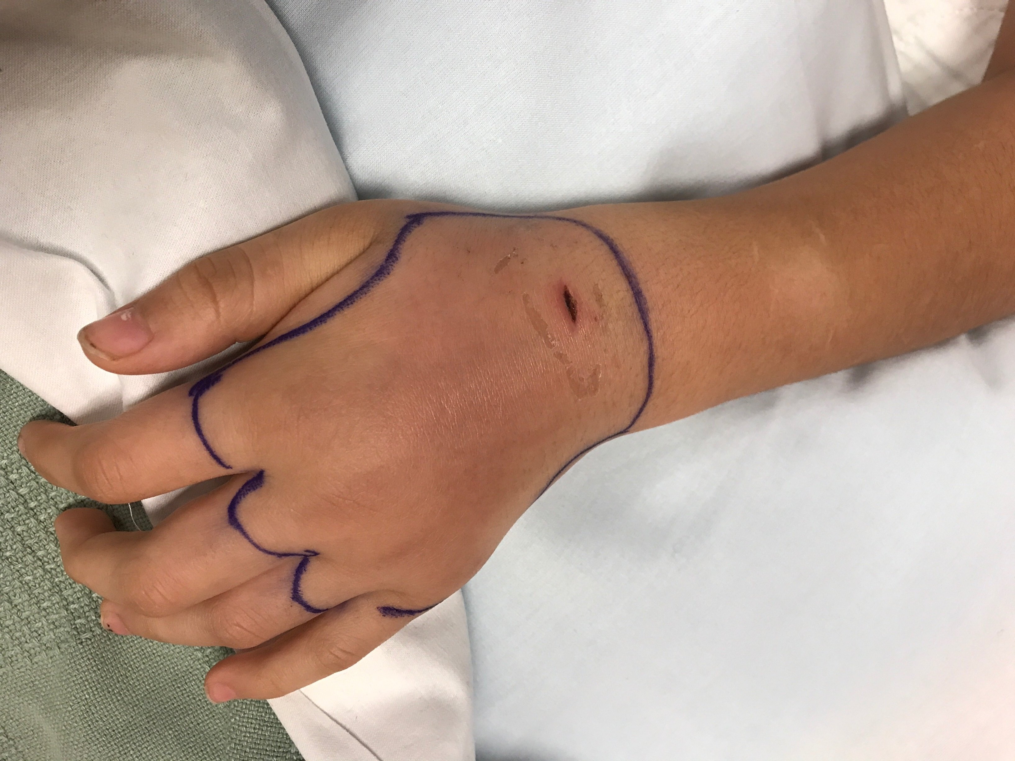
A 12-year-old female is admitted from the emergency department after failed outpatient therapy for a dog bite to her left hand. She was initially bitten by her family dog two days ago. The wound was appropriately cleaned with soap and water. The following day there was significant erythema and edema which prompted evaluation at an outside hospital. An X-ray of the hand did not show any foreign body and she was prescribed clindamycin prior to discharge. The swelling continued to extend beyond marker borders drawn the previous day and she continued to have intense pain and swelling. The patient experienced chills but did not have any fevers. She then presented to the emergency department due to worsening symptoms.
Her vital signs are unremarkable. The physical examination is notable for a 1 cm laceration on the dorsal surface of the hand/wrist without drainage. The erythema is mostly within the margins of the border drawn 24 hours earlier, but there is significant edema of the dorsal hand and wrist. Active range of motion is limited by pain, but she can flex her distal and proximal interphalangeal joints. There is significant tenderness over the dorsal surface of hand/wrist. Sensation to light touch is intact and capillary refill is <3 seconds on fingers. There is no epitrochlear or axillary lymphadenopathy. Initial white blood cell count is 7.95 x 10³ cells/mcL (58% neutrophils, no bands) with a CRP of 1.1 mg/dL.
Which of the following is the most appropriate antimicrobial regimen?
A. Ampicillin-sulbactam IV
B. Clindamycin IV
C. Vancomycin + ceftriaxone IV
D. Vancomycin + piperacillin-tazobactam IV
Answer: A. Ampicillin-sulbactam IV
Pasteurella species are the most frequent pathogens encountered in dog bites. Skin flora such as Streptococcus and Staphylococcus species can also be seen in addition to anaerobes such as Prevotella species. However, these infections are often polymicrobial and many of the anaerobic bacteria are beta-lactamase producers which need to be taken into consideration when selecting therapy.
The most appropriate treatment is ampicillin-sulbactam IV (amoxicillin-clavulanate if oral therapy is appropriate). This will adequately cover Pasteurella species, anaerobes, methicillin-sensitive Staphylococcus aureus (MSSA), and Streptococcus pyogenes. While lacking coverage for methicillin-resistant Staphylococcus aureus (MRSA), there are no historical clues or risk factors that indicate this is a likely pathogen.
Clindamycin is insufficient monotherapy for this patient and is considered a second-line treatment in appropriate combination. While it covers anaerobes and most skin flora, it does not adequately treat Pasteurella species and must be combined with a cephalosporin, trimethoprim-sulfamethoxazole (TMP/SMX), or a fluoroquinolone to provide appropriate coverage.
Vancomycin + ceftriaxone IV would provide appropriate coverage for MRSA and Pasteurella species. MRSA coverage would be excessive in this patient (no risk factors or history of MRSA infections provided) and there would be inadequate anaerobic coverage. Cephalosporin usage would require the addition of clindamycin or metronidazole.
Vancomycin + piperacillin-tazobactam IV is unnecessarily broad coverage for the above patient. While providing appropriate coverage for anaerobes, MRSA/MSSA, and Pasteurella species, this patient is not critically ill and there is no evidence of a necrotizing infection. Additionally, the synergistic effects of the antibiotics can increase risk for acute kidney injury.
Clinical Course
The patient’s blood cultures from admission grew Pasteurella multocida. Subsequent blood cultures showed no growth. While her erythema decreased and she did not have any leukocytosis or fever, she had worsening edema, increasing tenderness to palpation of her dorsal hand/wrist, and difficulty making a fist with passive range of motion. There was clinical concern for tenosynovitis and a deeper infection so magnetic resonance imaging (MRI) was obtained on hospital day 3. Imaging showed (1) a non-displaced metacarpal fracture, (2) metacarpal osteomyelitis, and (3) extensive tenosynovitis, myositis and cellulitis of the hand. There was fortunately no abscess present. Orthopedics performed irrigation and debridement of her wound on hospital day 4 with significant improvement in her symptoms. Four weeks of antibiotic treatment with extended-release amoxicillin-clavulanate was recommended by Infectious Diseases. She was discharged on hospital day 5 with significant improvement in her symptoms.
Clinical clues for P. multocida as a pathogen include quick (<24 hours) onset of swelling and pain and less extensive cellulitis. Staphylococcal and streptococcal infections have more intense cellulitis but take more time to develop. Pasteurella bacteremia (especially in a healthy patient) should prompt evaluation for a deeper infection (e.g., abscess, osteomyelitis, endocarditis).
References:
- American Academy of Pediatrics. Bite Wounds. Red Book: 2021-2024 Report of the Committee on Infectious Diseases: 169-175.
- Bula-Rudas and Olcott. Human and Animal Bites. Pediatr Rev. 2018; 39(10): 491-500.
- Stevens et al. Practice Guidelines for the Diagnosis and Management of Skin and Soft Tissue Infections: 2014 Update by the Infectious Diseases Society of America. Clin Infect Dis. 2014; 59: e10-52.